Force Microscopy
Researchers
Jeehoon Kim, Alex Frenzel
Instrument
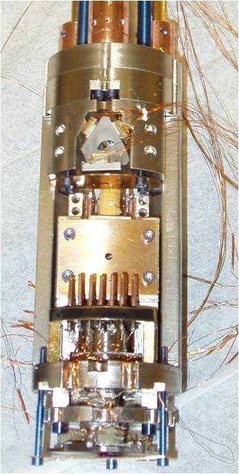
Fig. 1 Photo of force microscope.
We have constructed a custom variable temperature force microscope with the following features:
- Vibration isolation: floating room, floating table, internal suspension from damped springs.
- Temperature: 2K up to 340K
- Magnetic field: 5 Tesla vertical
- Scan Range: 35μm scan at room temperature;
several mm coarse (x ,y ,z ) motion of sample with respect to tip - Cantilever position detection:
fiber optic interferometer with λ=1550nm diode laser;
position noise better than 2×10⁻³ Å/√Hz - Geometry: allows either horizontal or vertical cantilever orientation

Fig. 2 Photo of force microscope setup, including vibration isolation.
Funding
NSF PHY 01-17795
Metal-Insulator Transition in VO2
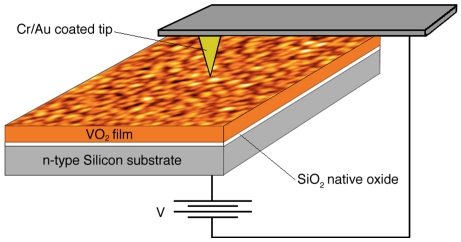
Fig. 3 Geometry of the atomic force microscope setup used to measure the conductivity of VO2 as a function of voltage applied to the sample.
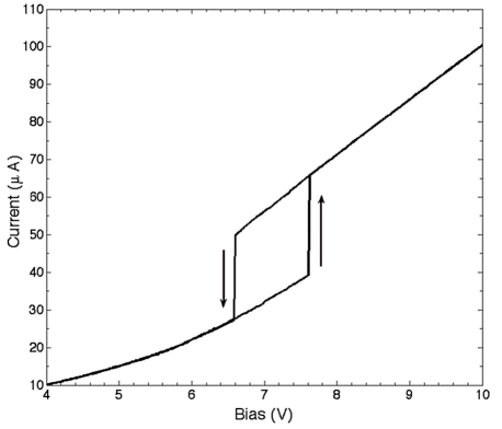
Fig. 4 Hysteresis loop in the conductivity of VO2 as a function of applied bias, in conducting-AFM geometry.
Collaborators
Shriram Ramanathan, Harvard University
Vortex Depinning in Iron Pnictide Superconductor NdO1-xFxFeAs
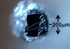
Fig. 5 Photo of single crystal NdO1-xFxFeAs sample, grown by the group of Prof. Paul Canfield, Ames Lab, Iowa.
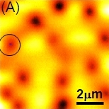
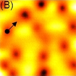
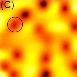
Fig. 6 We use the MFM to image and manipulate a vortex in NdO1-xFxFeAs at 5 K with a magnetic field of 1 Gauss. (A) Image at a "safe" height where tip-sample interactions are too weak to depin vortices. (B) Lower the tip to increase the interaction and drag a vortex. (C) Re-image at a "safe" height to demonstrate the vortex motion.
Collaborators
Paul Canfield, Ames Lab, Iowa